New and Novel Applications
Voltage-sensitive dyes are incredibly versatile and have shown great success in applications outside of the use of cardiac and neuronal cells. Recent publications show our dyes successfully being used to track electrical activity in human breast cancer cells and in live swine stomachs.
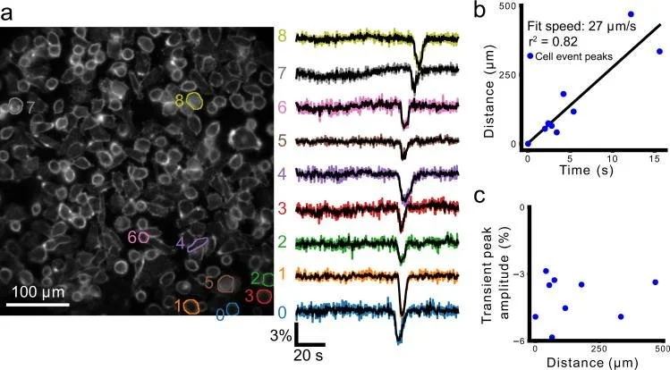
Quicke, Peter et al. “Voltage imaging reveals the dynamic electrical signatures of human breast cancer cells.” Communications biology vol. 5,1 1178. 11 Nov. 2022, doi:10.1038/s42003-022-04077-2
Ratiometric VSDs have been used in cancer cell lines to track hyperpolarization propagation in a population of cells (1). Regions of interest (ROIs) can be drawn around individual cells to measure fluorescence intensity changes as a function of time and distance from any chosen starting point. The speed of propagation can also be calculated.
ElectroFluor550™ (Di-4-AN(F)EP(F)PTEA) was used in these experiments, as it changes fluorescence in response to changes in membrane potential at a sub-microsecond speed. The ratiometric nature of this dye corrects for confounding factors such as uneven dye staining, photobleaching, and motion artifacts. This is particularly useful when investigating the propagation of hyperpolarization events.
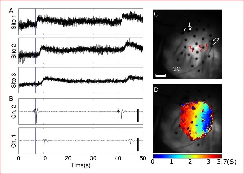
Zhang H, Yu H, Walcott GP, et al. High-resolution optical mapping of gastric slow wave propagation. Neurogastroenterol Motil. 2019;31(1):e13449. doi:10.1111/nmo.13449
Voltage-sensitive dyes are also being used in swine stomachs to map slow wave propagation in vivo. Methods have been developed to correct for motion artifacts, which can result in significant changes and invalidate the data collected.
Using farm pigs and staining in vivo with Di-4-ANEPPS resulted in the detection of depolarization propagation throughout a large area. This dye successfully stained pig stomachs despite active circulation in the live animal that could wash out the dye.
-
Quicke, P., Sun, Y., Arias-Garcia, M., Beykou, M., Acker, C. D., Djamgoz, M. B. A., Bakal, C., & Foust, A. J. (2022). Voltage imaging reveals the dynamic electrical signatures of human breast cancer cells. Communications biology, 5(1), 1178. Pubmed
Zhang, H., Yu, H., Walcott, G. P., Paskaranandavadivel, N., Cheng, L. K., O'Grady, G., & Rogers, J. M. (2019). High-resolution optical mapping of gastric slow wave propagation. Neurogastroenterology and motility, 31(1), e13449. Pubmed


