


ElectroFluor640™
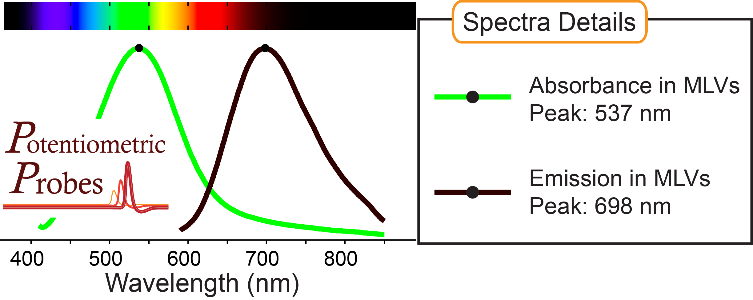
Absorbance, emission peaks in multilamellar lipid vesicles (MLVs) shown above: 538 nm, 696 nm
Absorbance peak in ethanol: 578 nm
Recommended excitation range: 630 nm - 650 nm
Compatible with ratiometric voltage imaging
MW: 562.74 g/mol
Other names: Di-4-ANEQ(F)BS (PP1015)
The minimum combined order total is $200
Highlighted Application
In this video, spontaneously beating human stem cell-derived cardiomyocytes stained with ElectroFluor640™ allows ratiometric optical action potential recordings. Propagation of electrical signals is also visible using this dye. The ratio of two excitation wavelengths boosts signals and removes artifacts due to non-uniform staining, bleaching, and the motion of these beating (contracting) human heart cells in a dish. Learn more on our Cardiac Research page.
Structure and Spectra

Product Details:
Pre-dried aliquots (solid, solvent removed)
Quantity: 100 nmol / tube (56 μg)
2.0-mL free standing polypropylene tubes
Sealed cap
Individually labeled