Structure and Spectra of TMRE
EP(F)PTEA from Potentiometric Probes")
EP(F)PTEA from Potentiometric Probes")
- Absorbance, emission peaks in multilamellar lipid vesicles (MLVs) shown above: 560.5 nm, 588 nm.
- Absorbance, emission peaks in ethanol: 551 nm, 578 nm.
- MW: 515.0 g/mol
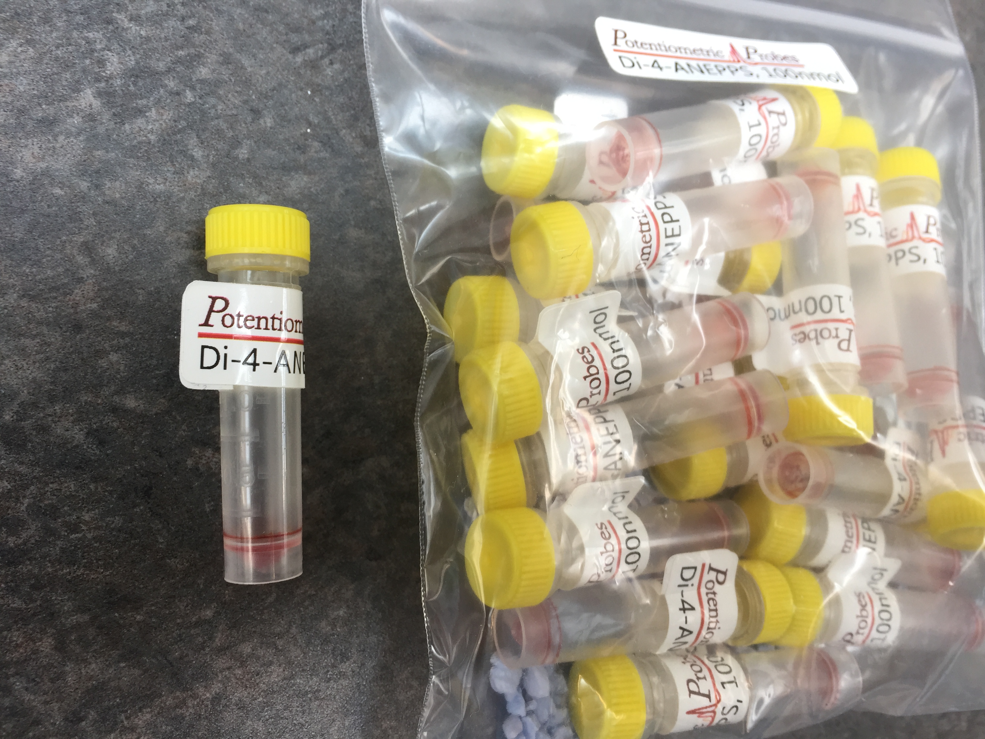
Unique Packaging for Convenience and Reliability
- Pre-dried aliquots (solid, solvent removed)
- Quantity: 100 nmol / tube (52 μg)
- 2.0-mL free standing polypropylene tubes
- Sealed cap
- Individually labeled
- Bagged with desiccant
- Bulk mg quantities also available
Order information
- See table for pricing
- Contact sales@potentiometricprobes.com for a quote
- Minimum order: 20. Price break: 100 tubes
- Shipping not included
- Other quantities and formats available upon request
- Inquire about “Academic Discount” for academic researchers
| TMRE – 100 nmol Pre-dried Aliquot Pricing | |||
|---|---|---|---|
| Product Number | Order Quantity (tubes) | Price / Tube | Order Total ($US) |
| 01790 | 20 | $4.00 | $80 |
| 01790 | 100 | $3.50 | $350 |
| TMRE, Bulk Pricing | |||
|---|---|---|---|
| Product Number | Order Quantity (mg) | Price / mg | Order Total ($US) |
| 01795 | 1 | $50 | $50 |
| 01796 | 5 | $40 | $200 |
Citations
- Ehrenberg, B., V. Montana, M. D. Wei, J. P. Wuskell, and L. M. Loew. 1988. Membrane potential can be determined in individual cells from the nernstian distribution of cationic dyes. Biophysical journal 53:785-794. PubMed
- Farkas, D. L., M. D. Wei, P. Febbroriello, J. H. Carson, and L. M. Loew. 1989. Simultaneous imaging of cell and mitochondrial membrane potentials. Biophysical journal 56:1053-1069. PubMed
- Loew, L. M., R. A. Tuft, W. Carrington, and F. S. Fay. 1993. Imaging in five dimensions: time-dependent membrane potentials in individual mitochondria. Biophysical journal 65:2396-2407. PubMed
- Loew, L. M., W. Carrington, R. A. Tuft, and F. S. Fay. 1994. Physiological cytosolic Ca2+ transients evoke concurrent mitochondrial depolarizations. Proceedings of the National Academy of Sciences of the United States of America
- Fink, C., F. Morgan, and L. M. Loew. 1998. Intracellular fluorescent probe concentrations by confocal microscopy. Biophysical journal 75:1648-1658. PubMed
- Ward, M. W., A. C. Rego, B. G. Frenguelli, and D. G. Nicholls. 2000. Mitochondrial membrane potential and glutamate excitotoxicity in cultured cerebellar granule cells. Journal of Neuroscience 20:7208-7219. PubMed
- Creed, S., and M. McKenzie. 2019. Measurement of Mitochondrial Membrane Potential with the Fluorescent Dye Tetramethylrhodamine Methyl Ester (TMRM). In Cancer Metabolism: Methods and Protocols. M. Haznadar, editor. Springer New York, New York, NY. PubMed
- Perry, S. W., J. P. Norman, J. Barbieri, E. B. Brown, and H. A. Gelbard. 2011. Mitochondrial membrane potential probes and the proton gradient: a practical usage guide. BioTechniques 50:98-115. PubMed
- Chazotte, B. 2011. Labeling Mitochondria with TMRM or TMRE. Cold Spring Harbor Protocols 2011. PubMed
- Crowley, L. C., M. E. Christensen, and N. J. Waterhouse. 2016. Measuring Mitochondrial Transmembrane Potential by TMRE Staining. Cold Spring Harbor Protocols 2016. PubMed